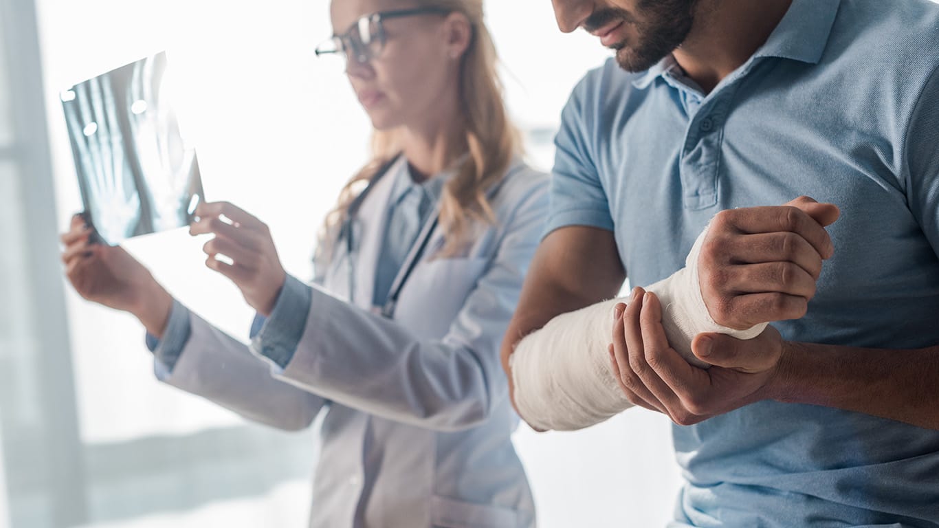
Shoulder pain and reduced mobility are common complaints among both physically active and sedentary adults, especially during middle age. The shoulder is the most mobile joint in the human body, with a complex architecture and multi-planar range of motion. Optimal shoulder mobility depends on the harmonious interaction of various tissues and structures that surround and act upon the shoulder, and shoulder pain can arise from multiple sources.
It is not enough to treat shoulder pain with drugs and steroid injections, and shoulder surgery poses multiple risks, with no guarantee of success. To fully restore functional shoulder mobility, you need to address the underlying causes of pain and dysfunction. Learn how new technologies and orthobiologic solutions are changing the game for shoulder rehabilitation.
FOOD NEWS: 10 celebrity chef restaurants to try in Arizona
THINGS TO DO: Want more news like this? Get our free newsletter here
Factors that Influence Shoulder Mobility and Stability
Thanks to its ball-and-socket architecture, the shoulder is the most mobile joint in the human body, enabling the arm to move freely in all three planes of motion. However, the price we pay for shoulder mobility is reduced joint stability, making the glenohumeral joint and its surrounding structures more vulnerable to injury.
Some factors that influence shoulder stability include:
- The glenoid labrum, the fibrocartilaginous structure that lines the glenoid fossa, increases the contact area of the head of the humerus, keeping it from disengaging from the socket.
- The glenohumeral and coracohumeral ligaments reinforce the joint capsule and hold the humeral head in place. Overly tight ligaments can limit mobility, while overly lax ligaments reduce joint stability.
- Bony structures – particularly the humeral head and the glenoid fossa – must fit together to allow for optimal range of motion. Stability is reduced by a shallow glenoid or abnormal bone shapes. When the humerus fits snugly in the glenoid fossa, it produces a slight vacuum effect that helps to hold the humeral head in place.
- Rotator cuff muscles and their tendons govern and control shoulder movement. The four rotator muscles press the humeral head into the glenoid, creating a concavity-compression mechanism that resists shoulder translation. Imbalances of the rotator cuff produced by muscle tightness or weakness reduce shoulder stability and function.
- The scapular stabilizer muscles – the trapezius, serratus anterior, and rhomboids – control positioning and movement of the scapula (shoulder blade), and promote proper alignment of the glenoid during arm movement. Scapular dyskinesis is strongly linked to shoulder instability.
- The deltoid and biceps brachii muscles of the arm provide additional compressive forces that control and stabilize the shoulder.
Shoulder stability relies on neuromuscular control and coordinated muscle activation patterns, along with balanced activation of opposing muscle groups. Fascia works together with muscles to provide biotensegrity – elastic tension that guides and controls movement, and mediates internal and external forces.
Traumatic injuries, repetitive overuse, and muscle disuse can all affect shoulder mechanics and undermine biotensegrity, causing shoulder instability. Poor posture creates muscle imbalances in the rotator cuff and shoulder girdle that destabilize the shoulder and increase injury risk. Age-related degeneration of the shoulder complex reduces stability over time.
Common Types of Shoulder Dysfunction
Clearly, many factors play into shoulder mechanics, and shoulder pain and dysfunction can arise spontaneously from overuse, disuse or misuse of the surrounding tissues and structures. During shoulder movement, muscles, tendons, ligaments, fascia, bones, nerves and blood vessels all work interdependently, and all must interact in harmony.
Common types of shoulder conditions include:
- Rotator cuff lesions, ruptures and tendinopathy
- Shoulder bursitis
- Impingement of tendons, bursae, nerves and blood vessels by other structures
- Labral cartilage ruptures
- Shoulder dislocation
- Joint hypermobility leading to shoulder instability
- Frozen shoulder syndrome (adhesive capsulitis)
- Shoulder osteoarthritis
- Scapular pain and dyskinesis
- Fascial densifications and adhesions
Treating the locus of shoulder pain without differentiating the underlying cause(s) can result in inappropriate or inadequate treatment that fails to provide shoulder stability and restore pain-free functional mobility.
Rehab Success Relies on Accurate Diagnosis
Conventional shoulder pain diagnosis typically involves a review of health history and a clinical exam that includes testing for shoulder strength and range of motion, and special tests to identify conditions like rotator cuff tears, impingement, or instability. Your doctor will check for signs of swelling, deformity, or muscle atrophy.
Further tests with arthroscopy and nerve studies may be recommended. Imaging by X-ray can reveal bone fractures or deformities, and MRI may be prescribed to evaluate soft tissues. However, those imaging methodologies give only limited insight, and they involve exposure to radiation, which poses safety risks for some patients.
For integrative and holistic practitioners, high-resolution ultrasonography is becoming the imaging mode of choice. Not only is ultrasound imaging safe for almost all patients, but it is inexpensive compared to MRI, and delivers immediate results in real time.
High-resolution diagnostic ultrasound empowers clinicians to:
- Compare the injured and uninjured shoulders
- View nerves, muscles, fascia and bones along their entire path
- Visualize the interaction of various structures during dynamic movement
- Identify fascial densifications and adhesions, and myofascial trigger points
- Pinpoint the exact site of nerve compression or entrapment
- View multiple tissues and structures in a single session
- Elicit patient feedback during the imaging session
- Leverage sonoelastography to test tissue density
- Access superb microvascular imaging to detect early signs of tissue regeneration
High-resolution ultrasound provides an advanced tool for comprehensive diagnosis, ensuring that nothing is overlooked. It dramatically reduces the risk of being under-diagnosed or misdiagnosed, increasing your chances of successful treatment.
New smart technologies are also gaining popularity among holistic practitioners whose patients want to restore mobility without the use of drugs or surgery. Such advanced devices take the guesswork of assessment and diagnosis, and provide objective data to help guide and measure the rehabilitation process.
ShowMotion™ is an objective tool for analyzing shoulder movement using motion tracking sensors, placed on the patient’s skin to collect data about movement quality. The patient performs a series of joint-specific movements, and the data is analyzed by ShowMotion’s proprietary software, providing valuable insights about inefficient muscle coordination patterns that reduce motor efficiency.
The Neuralign Shoulder Pacemaker is a shoulder rehabilitation device with a kinematic sensor activated by movement. The patient dynamically interacts with the device to emulate efficient muscle recruitment patterns, enhance movement quality, and restore optimal muscle balance during rehabilitation.
Ultrasound-Guided Regenerative and Manual Therapies Restore Biotensegrity
Multimodal Shockwave Therapy
Ultrasound guided extracorporeal shockwave therapy (ESWT) delivers radial, linear, focused and defocused shockwaves to address different tissue types. The high frequency acoustic waves relieve pain, reduce inflammation, and have a regenerative effect on damaged tissues. ESWT helps to realign collagen fibers, promotes hydration of fascial tissues, and restores tissue gliding.
Ultrasound-Guided Dry Needling
Dry needling addresses myofascial trigger points – hard knots of tightly contracted fibers that cause pain and disrupt biotensegrity. During the procedure, filament-thin non-medicated needles are inserted through the skin into the trigger point, causing a twitch response that immediately relaxes the fibers. Research published in 2023 by Kalika et al. found that dry needling of mechanically obstructed shoulder structures, particularly the acromioclavicular joint (ACJ), resulted in improved shoulder mobility and decreased myofascial pain.
PENS
Percutaneous neuromodulation (PENS) is a therapeutic approach that uses electrical stimuli to calm and desensitize hyperactivated nerves. It involves the insertion of several filament-thin needles under ultrasound guidance into muscle tissue adjacent to the targeted nerve. PENS stimulates the nerve with varying waves of low frequency electrical current to help restore optimal neural function.
Stecco Myofascial Release
The Stecco method of fascial manipulation involves deep friction that heats up tissues and stimulates mechanical action. Stecco fascial manipulation is an evidence-based methodology for breaking up scar tissue and releasing adhesions, to restore the integrity of fascial tissue. Patients often report immediate pain relief after a single Stecco session.
Orthobiologics: A New Frontier in Shoulder Rehab
Orthobiologic injection therapies use natural/neutral solutions, injected with precision under ultrasound guidance. The injected solutions stimulate cellular repair by either nourishing or irritating the targeted cells. Injections are also invaluable for liberating nerves and blood vessels entrapped by densified fascia, to restore gliding.
Platelet Rich Plasma (PRP)
PRP therapy extracts a high concentration of platelets from a sample of the patient’s own whole blood. When injected into damaged tissues, PRP initiates tissue repair by releasing biologically active agents such as growth factors, cytokines, lysosomes and adhesion proteins.
Matrix PRP
For tendon ruptures, Matrix takes PRP therapy to the next level by creating a collagenous bridge between the walls of the tear and the rest of the tendon. Matrix is a highly concentrated PRP, diluted and mixed with fibrinogen. At the injection site, the solution becomes a gel-like collagenous substance that adheres to the walls of the tear and fills the space between them, creating a fibrin matrix that helps to stabilize growth factors and attract stem cells to the treatment site.
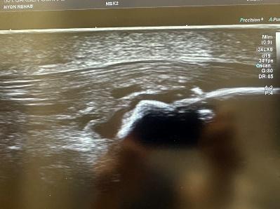
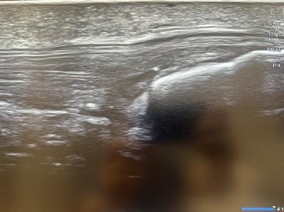
Tendon tear before and after Matrix PRP
Platelet Releasate Therapy
Platelet releasate therapy involves injecting platelet releasate – a mixture of growth factors and biomolecules – into injured muscles and tendons. The solution activates leukocytes and endothelial cells, and stimulates blood vessel growth, to increase the flow of oxygen, nutrients and growth factors to the damaged tissues.
Alpha-2-Macroglobulin (A2M)
Alpha 2 macroglobulin (A2M) is a naturally occurring blood plasma protein that acts as a carrier for numerous proteins and growth factors. As a protease inhibitor, A2M reduces inflammation in arthritic joints and helps to deactivate a variety of proteinases that typically degrade cartilage.
Prolotherapy and Prolozone
Prolotherapy uses a biologically neutral solution to irritate stubborn tissues, triggering the body’s innate healing mechanisms to grow new normal tendon, ligament and muscle fibers. Prolotherapy is often used for slow-to-heal tendon and ligament ruptures, where low vascularity inhibits tissue healing.
Prolozone takes Prolotherapy to the next level by adding a combination of procaine, anti-inflammatory medications, vitamins, and minerals, followed by a mixture of ozone/oxygen gas, injected into targeted joints or tissues. When performed under ultrasound guidance, Prolozone therapy quickly reduces pain and inflammation while jump-starting the healing process.
Hyaluronic Acid Injections
Hyaluronic acid is a natural component of joint synovial fluid, and a key component of fascia. Its slippery gel-like properties provide lubrication that reduces friction, enabling joints, muscles and fascia to move freely without pain. Hyaluronic acid injections can help to reduce friction in arthritic joints, and revitalize fascia’s functional properties.
Interfascial Plane and Nerve Hydrodissection
Shoulder pain often involves damaged fascial tissue that has thickened and become sticky, often adhering to other structures. The hydrodissection procedure injects a saline solution into densified fascial layers under ultrasound guidance, separating the layers and releasing entrapped nerves and blood vessels.
Skip the Scalpel with Holistic Shoulder Treatment
Numerous sports, occupations and everyday activities depend on a strong, stable, and mobile shoulder complex. Treating shoulder pain symptoms without addressing their underlying causes can trap you in an endless spiral of injury-recovery-reinjury that keeps you in a state of perpetual shoulder pain until surgery becomes your only option.
Holistic therapy halts the spiral by healing damaged tissues and restoring biotensegrity. Advanced technologies and therapies support and accelerate the healing process, putting an end to shoulder pain and dysfunction so you can get back to doing the things you love.
Author: Dr. Lev Kalika, DC, clinical director of NYDNRehab, is an internationally recognized expert in diagnostic and musculoskeletal ultrasonography, with multiple research papers to his credit. Dr. Kalika has studied with some of the world’s most prestigious experts in diagnostic, fascia, and nerve ultrasonography, and has presented his research at multiple international conferences. Dr. Kalika studied directly under Dr. Ben Kibler, world-renowned orthopedic surgeon and pioneer of scapular dyskinesis. For orthobiologic procedures, Dr. Kalika partners with Dr. Yuri Brosgol, MD, a seasoned expert in treating myofascial pain.